Nederlands
AI heart measurement predicts risk of death slightly better than doctors' measurement
An AI program uses an MRI scan to measure how well the heart pumps, in a few seconds. In 27,382 patients, that measurement was slightly more strongly linked to their chance of survival than the measurement by experienced doctors. A doctor still checks the results.
How well does the heart pump? That is one of the most important questions in cardiology. Doctors measure it with the ejection fraction: the share of blood that the left ventricle, the large pumping chamber of the heart, pushes out with each beat. That number helps decide whether someone gets medicines for heart failure, a small device in the chest such as a pacemaker, or a procedure that improves the blood supply to the heart. Doctors also keep an eye on it during chemotherapy, which can damage the heart.
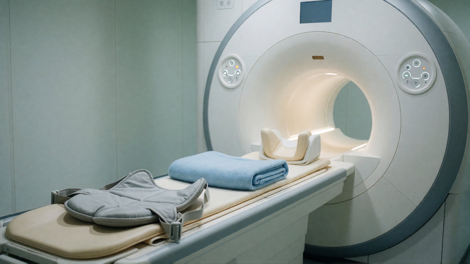
The most accurate measurement comes from an MRI scan of the heart: a scanner that uses a strong magnet and radio waves to make moving images of the heart. A specialist traces the edge of the heart chamber on those images. According to earlier work by the same research team, that takes about thirteen minutes per patient.
Seconds instead of minutes
Researchers at City St George's, University of London and University College London (UCL) had an AI program do the same work. The program was developed by Rhodri Davies, associate professor at UCL and one of the leaders of the study. It needed two to three seconds per patient.
The team compared the measurements by the program and by experienced doctors in 27,382 patients from the United Kingdom and the United States. These patients had an MRI scan because they had a heart condition or were suspected of having one. They were then followed for an average of more than five years. During that time, 3,458 of them died. The study was published in the medical journal The Lancet Digital Health.
The question was which measurement gives the best estimate of who is at greater risk of dying. That turned out to be the one by the AI program: a small difference, but larger than chance can explain. This stayed the case when the researchers took age and sex into account, although the gain then became smaller. According to Davies, such a small improvement does count, because it concerns thousands of patients.
A doctor still checks
The program made a mistake in about 1 per cent of the patients, affecting 0.08 per cent of all images. That is why a doctor must keep checking the results and assess the scan together with everything else that is known about the patient. The team sees the program as a first measurement, with a specialist checking where needed.
The study looked back at existing scans. It shows which measurement is more closely linked to death, but not yet whether patients live longer when their doctor uses the AI measurement. Differences in how doctors trace the heart chamber may also explain part of the difference. It has not been disclosed when the scans were made.
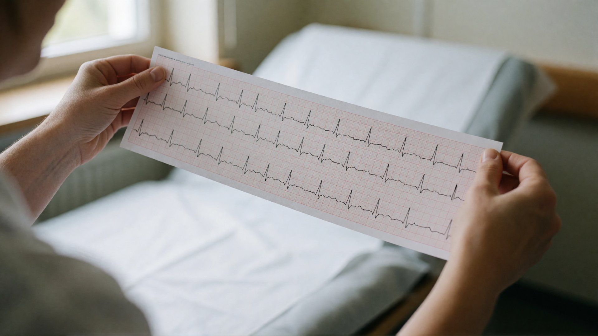
The researchers now want to combine the measurements from heart scans with other tests, especially the ECG, a simple recording of the heart's electrical activity. In this way they hope to find problems in the heart muscle earlier, so that treatment can start before the heart is permanently damaged.
What does this mean for the road to the singularity?
Computers that read medical images are no longer new. What is new is that a large study has checked whether the measurement by an AI program also says something about how patients fare years later. If a measurement that now takes thirteen minutes of specialist work takes a few seconds, a hospital can measure every heart scan, including old scans from the archive. The specialist then has more time for the difficult cases and for talking to the patient.
If that trend continues, a second advantage follows. Measurements that are quick, cheap and always done the same way are easy for researchers to combine with other data. Another AI model has already found a previously unknown pattern in ordinary ECGs that is linked to sudden cardiac death. Hospitals will need to keep checking whether such a program also works for patients and scanners it has not seen before. If that succeeds, a doctor may be able to combine signals from the ECG and the heart scan to see a heart problem coming while treatment can still prevent a lot.
What is the technological singularity? Read the explanation →