Nederlands
Anthropic uses AI to determine, for the first time, the shape of a puzzling protein shell from the bacterial world
Anthropic's biology team has used its AI assistant Claude to determine, for the first time, the 3D shape of a vault from the bacterial world: a hollow protein shell made with a protein from a bacterial virus. What such shells do in cells is now being studied further.
In the cells of humans and many other organisms with complex cells, there are large, hollow protein shells whose purpose nobody knows for certain. They are called vaults: their ribbed shape reminded their discoverers of the ceiling of a cathedral.
The biology team at the AI company Anthropic has now used its AI assistant Claude to determine, for the first time, the 3D shape of a vault from single-celled organisms without a cell nucleus, such as bacteria. The team made the shell in E. coli, a widely used laboratory bacterium, using a protein from a bacteriophage: a virus that infects bacteria. Anthropic announced the results on 4 November 2026.
Forty years of mystery
Leonard Rome and Nancy Kedersha discovered vaults in 1986. In mammals, the shell consists of 78 copies of the same protein, split into two identical halves. Researchers have suggested that vaults carry molecules between the cell nucleus and the rest of the cell, protect cells against toxic substances or support the immune defence. None of these ideas has been confirmed. Mice without vaults live normally, which may mean that vaults are only needed under certain conditions.
Searching 15 billion proteins
The team looked for relatives of the vault protein in simpler organisms. Using Claude Science, Anthropic's working environment for researchers, it searched a database of about 15 billion predicted proteins. Relatives turned out to occur in 29 large groups of bacteria and four large groups of archaea: single-celled organisms without a cell nucleus that resemble bacteria but form a branch of life of their own. Even so, they were found in fewer than 1 per cent of the species whose genetic material is known.
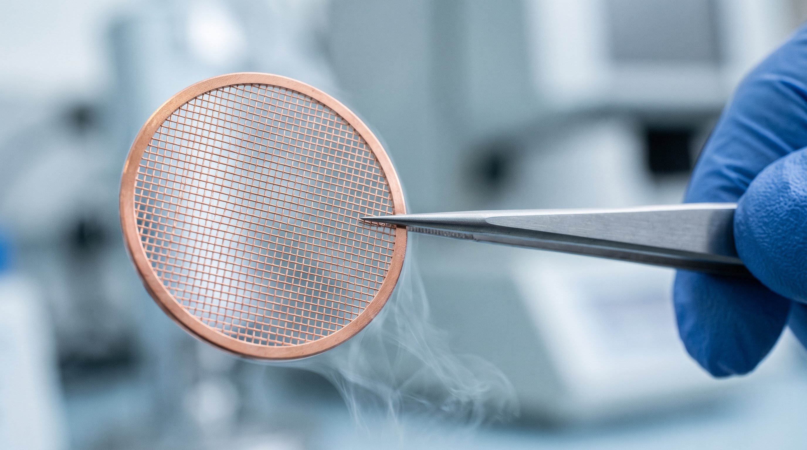
Of 44 chosen proteins, the researchers first tested in a test tube, without living cells, which ones were produced well. The best 24 went on to E. coli. For 15 of them, the protein stayed neatly dissolved instead of clumping together. Seven of those 15 formed shells that looked like vaults. Using cryo-electron microscopy, the team confirmed the shape of four: two with proteins from bacteria and two with proteins from bacteriophages. In this technique, samples are frozen very rapidly and photographed with an electron beam; from thousands of images of individual shells, the computer calculates a 3D image.
Rounder and roomier
For the detailed study, the team chose a protein from a bacteriophage, because it formed many complete, closed shells. This shell consists of 96 copies of the same protein, compared with 78 in mammals. It is about 54 nanometres long and 45 nanometres wide; more than 20,000 of these shells fit side by side across a millimetre. A mammalian vault measures about 67 by 40 nanometres. The new shell is therefore shorter but wider, and has flatter ends. As a result, according to Anthropic, it encloses about 25 per cent more space.
Each protein has a small cavity with a small molecule inside that has not yet been identified. Claude noticed that its shape resembles coenzyme A, a common helper molecule in cells. This is a lead for further research, not yet a confirmation.
The team also made 15 modified versions of the protein, mostly with changes at the places where neighbouring proteins touch. Some changes near the ends meant that the shell no longer closed: open particles in the shape of a rosette formed instead. This shows that exactly that part is needed to close the shell.
The part of the protein that forms the wall consists of seven repeated segments; in mammals there are nine. When the researchers doubled two segments, bringing the number to nine, a larger shell formed with about 20 per cent more interior space. According to the researchers, this may explain how vaults changed in size during evolution. They also think of applications, such as tiny capsules that deliver enzymes. That is a next step that still has to be investigated.
This is a different way of working from Microsoft's BioEmu-2, an AI model that predicts which shapes a protein can take. There, AI calculates the shape. Here, the shape was measured with the microscope, and Claude helped with the experiments and with processing the images.
What Claude did
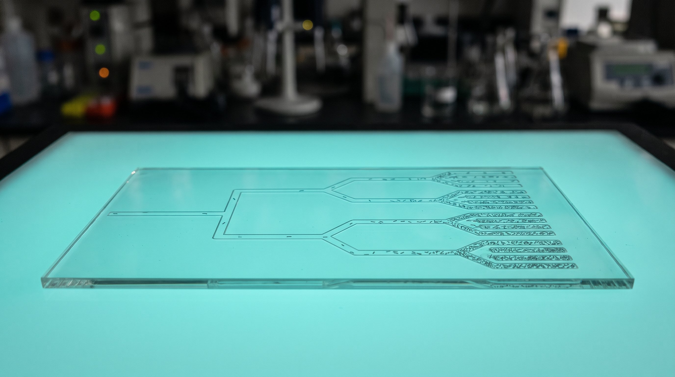
Claude helped with searching for proteins, designing DNA and purifying the shells: separating them from the rest of the cell. The usual method, in which the particles are sorted in a centrifuge through a sugar solution, takes about three days. The team instead let the cell contents run through a tube filled with porous beads. Small molecules wander through the pores and leave the tube later, while large shells run through quickly. In this way, a sample was ready for the microscope in about 90 minutes.
Claude was at first against this approach, because clumped protein leaves the tube at the same time and so cannot be separated from the shells. When the team went ahead anyway, Claude recommended a suitable tube and suggested putting the contents of broken-open cells straight into the tube. The team hesitated because of the cost and the risk of clogging, but it worked.
For the analysis of the microscope images, Claude directed the processing itself, using the freely available program RELION. The researchers advised on the choices. A first complete model of the structure was ready after 34 hours, and the final 3D images after about a week. According to the team, this work normally takes weeks. That analysis did use hundreds of graphics processors at once, more computing power than many universities have. According to Anthropic, most of the other work can also be done in an ordinary lab, with the existing Claude products. The whole project took less than four months.
Mistakes and lessons
Claude also made mistakes. A work protocol written by Claude contained an additive in the growth medium that hindered the uptake of DNA by the bacteria. This cost the project about two weeks. In an early analysis, Claude skipped a standard step, which produced a 3D image mixed with its own mirror image. The researchers then asked Claude not to improvise any more and to follow the standard procedures unless they do not work.
The team shared its lab notes with the researchers who train Claude. According to Anthropic, Claude got better at these tasks as newer models were released during the project.
What is still being investigated
So far, the shells have only been made in E. coli. The team will test whether they also form in the bacteria and bacteriophages where the proteins occur naturally, and what they do there. Leonard Rome, who advised the team and was paid for it, calls finding that function an enormous challenge. He considers it possible that vaults have the same task everywhere, but also that they have taken on different tasks in different organisms.
The research report has been submitted to bioRxiv, a platform for scientific papers that have not yet been reviewed by other experts. Anthropic plans to release the 3D images and more data soon, so that other researchers can build on them.
What does this mean for the road to the singularity?
This is an example of AI taking part in almost every step of a biological experiment, from searching 15 billion proteins to processing microscope images. A first 3D model in 34 hours, where the team would normally calculate for weeks, gives an idea of how much faster basic research can become. The mistakes show that the researchers keep steering the work. At the same time, according to Anthropic, Claude improved during this very project as newer versions appeared. Because most of the work can also be done in an ordinary lab, more research groups may soon work this way. The next step is to find out what these shells do in nature and whether they can be used as tiny capsules.
What is the technological singularity? Read the explanation →